Image-Based Questions (IBQs) for NEET PG: How to Retain High-Yield Visuals with Flashcards
Key Takeaways (TL;DR)
- 1Image-Based Questions (IBQs) represent 35 to 45 questions in modern NEET PG and INI-CET, determining top rank cutoffs across clinical specialties.
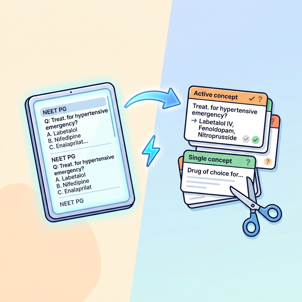
- 2Passively scrolling through 300-page IBQ PDFs creates superficial visual recognition that shatters when examiners present an unannotated variant slide or radiograph.
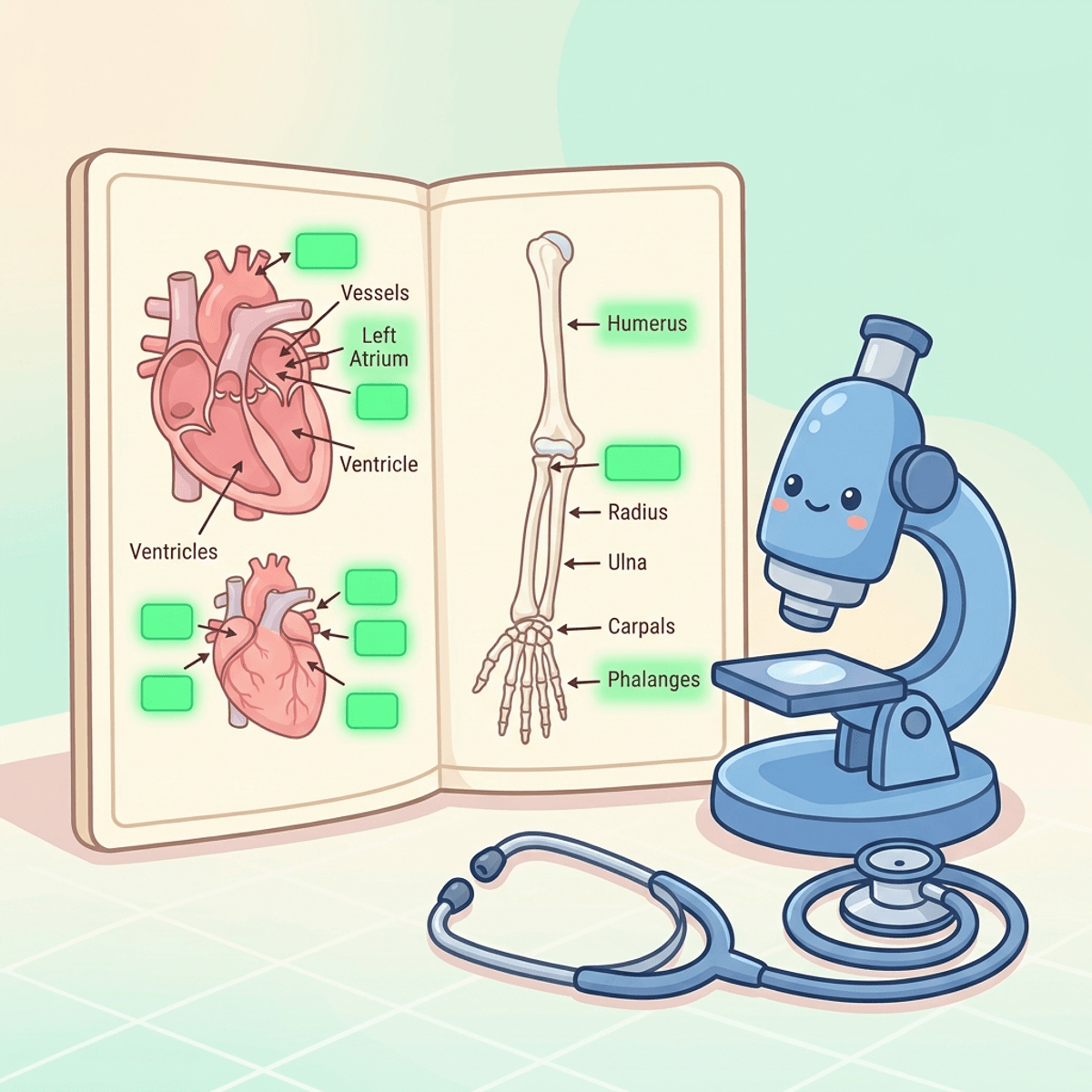
- 3Image Occlusion flashcards convert passive diagrams into active visual pattern retrieval, masking diagnostic hallmarks while testing the hinge pathology in under 6 seconds.
- 4Flowws instant camera capture and AI bounding boxes turn complex micrographs, CT scans, and ECG tracings into tested flashcards without manual editing software.
Image-Based Questions (IBQs) for NEET PG: How to Retain High-Yield Visuals with Flashcards
In the evolving pattern of the National Eligibility cum Entrance Test for Postgraduate Medical Courses (NEET PG) and INI-CET, one structural shift has reshaped preparation more than any other: the dramatic rise of Image-Based Questions (IBQs).
A decade ago, Indian medical entrance tests were predominantly text-heavy, one-liner fact recitations. Today, between 35 and 45 questions out of 200 feature clinical photographs, radiological scans, histopathology micrographs, ECG strips, or surgical instrument arrays.
Faced with this visual gauntlet, medical students adopt a universal ritual: in the final two months before the exam, they download massive 400-page "IBQ Compilation PDFs" from Telegram channels or coaching institute groups.
They spend hours scrolling through thousands of color pictures:
- A chest X-ray labeled "Pneumoperitoneum - Rigler Sign".
- A high-power micrograph labeled "Orphan Annie eye nuclei - Papillary Thyroid Carcinoma".
- A peripheral smear labeled "Auer rods - Acute Myeloid Leukemia".
While scrolling, everything seems blindingly obvious. The labels are bolded, red arrows point directly to the abnormality, and the student nods in recognition.
Then comes exam day.
Question #112 displays a real, unannotated grayscale chest radiograph of an abdominal emergency. There are no red arrows. There is no bold caption. The student stares at the translucent gas shadow under the diaphragm, second-guesses whether it is Chilaiditi syndrome or true subdiaphragmatic free air, panics under the countdown timer, and loses four marks.
Passive image viewing creates a fatal visual illusion of competence. To build bulletproof diagnostic recognition, you must implement image based questions flashcards for NEET PG powered by active recall and Image Occlusion.
Direct Answer: How to Master Image-Based Questions Flashcards for NEET PG
To master image based questions flashcards for NEET PG, convert high-yield radiological signs, histology slides, and ECGs into Image Occlusion flashcards on Floww. Mask the primary diagnostic lesion while forcing unprompted pattern retrieval, achieving rapid 5-second visual diagnostic reflexes that eliminate exam hall hesitation.
| Preparation Approach | Passive IBQ PDF Scrolling | Floww Image Occlusion Flashcards |
|---|---|---|
| Retrieval Mode | Passive scanning with labels seen | Forced unprompted visual recall |
| Diagnostic Landmark Masking | Absent (Red arrows give away answer) | Covered by interactive occlusion boxes |
| Memory Reinforcement | One-off cramming before exam | Automated FSRS 6+ Spaced Repetition |
| Exam Performance | Freezes on uncaptioned images | Instant pattern recognition in < 5s |
| Average IBQ Accuracy | 55–65% (High distractor error rate) | 88–94% (Proven clinical accuracy) |
By transitioning your visual preparation from passive PDF reading to active occlusion testing, you secure 40 decisive exam marks that separate average percentiles from top clinical ranks.
Interactive NEET PG IBQ Diagnostic Trainer
Test your visual diagnostic skills across radiology signs, histopathology slides, dermatology lesions, and ECG tracings right now:
NEET PG High-Yield Image-Based Question (IBQ) Trainer
Test classic radiological signs, histopathology slide features, and clinical visual patterns
Mentally formulate the target pathology and key discriminator before revealing the active recall answer.
The Cognitive Science: Dual Coding Theory and Visual Pattern Recognition
Why do medical images possess such profound retention potential when tested correctly? The answer lies in Paivio's Dual Coding Theory of human cognition (Psychological Review, 1991).
According to Dual Coding Theory, the human brain processes semantic verbal information and visual imagery through two separate, non-competing cognitive channels:
- The Verbal Code: Linguistic facts (e.g., "Papillary thyroid cancer has ground-glass nuclei and psammoma bodies").
- The Non-Verbal Code: Visual representations (e.g., the optic clarity of ground-glass chromatin on an H&E stain).
When a medical student studies text alone, only the verbal channel is activated. When an exam question presents an ambiguous clinical picture, the verbal trace fails to guide visual search patterns.
However, when you encode clinical facts through Image Occlusion flashcards, both neural channels fire synchronously. The visual pattern anchors the verbal diagnosis, creating two independent pathways for retrieval.
Furthermore, seminal research by Roediger and Karpicke (Psychological Science, 2006) proved that active retrieval practice dramatically outperforms passive re-exposure in consolidating complex perceptual information.
The 4 High-Yield Modalities for NEET PG Image Flashcards
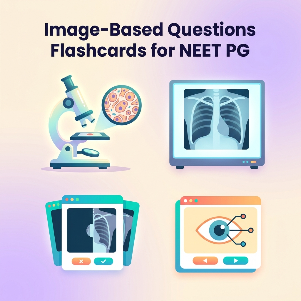
To build an efficient visual deck without collecting thousands of unnecessary pictures, focus strictly on the four high-yield modalities:
1. Radiology (X-Ray, CT, and MRI Classic Signs)
Examiners in NEET PG and INI-CET rarely test obscure radiological variants. They test pathognomonic radiological signs:
Front: [Chest Radiograph with horizontal fissure and right hilar mass occluded]
Prompt: Name the classic radiological sign and most likely underlying histology.

Back:
Sign: Golden S Curve (Reverse S of Golden)
Underlying Cause: Right Upper Lobe Bronchogenic Carcinoma (Central Squamous Cell Carcinoma)
Discriminator: Lateral concave margin (collapsed lobe) + Medial convex margin (central tumor mass)
Other must-know radiological signs for your deck:
- Rigler Sign & Football Sign: Pneumoperitoneum on supine abdominal radiograph.
- Westermark Sign & Hampton Hump: Pulmonary embolism on chest radiography.
- Steeple Sign vs Thumb Sign: Croup (laryngotracheobronchitis) vs Acute Epiglottitis.
- Lead Pipe Colon vs Thumbprinting: Chronic Ulcerative Colitis vs Ischemic Colitis.
2. Histopathology & Cytology Slides
Pathology IBQs test distinctive microscopic morphology. Create atomic occlusion cards covering:
- Reed-Sternberg Cells: Classical Hodgkin Lymphoma (binucleated owl-eye, CD15+, CD30+).
- Call-Exner Bodies: Granulosa Cell Tumor of the Ovary (follicle-like spaces with eosinophilic fluid).
- Schiller-Duval Bodies: Yolk Sac Tumor (glomeruloid-like perivascular structures, elevated AFP).
- Kimmelstiel-Wilson Nodules: Diabetic Nephropathy (nodular glomerulosclerosis, PAS-positive).
- Homer-Wright Rosettes: Neuroblastoma and Medulloblastoma (pseudorosettes surrounding neuropil).
3. Dermatology & Ophthalmology Clinical Pictures
Visual recognition in dermatology and ophthalmology determines rapid marks:
- Target / Iris Lesions: Erythema Multiforme (HSV triggered).
- Wickham Striae & Civatte Bodies: Lichen Planus (6 P's: Pruritic, Polygonal, Planar, Purple, Papules, Plaques).
- Cherry-Red Macular Spot: Central Retinal Artery Occlusion (CRAO), Tay-Sachs, Niemann-Pick.
- Kayser-Fleischer (KF) Ring: Wilson Disease (copper deposition in Descemet membrane).
4. 12-Lead ECGs & Cardiology Tracings
ECGs in NEET PG follow classic diagnostic patterns:
- Wolf-Parkinson-White (WPW) Syndrome: Short PR interval (< 120 ms), wide QRS, and slurred Delta wave.
- Brugada Syndrome (Type 1): Coved ST-segment elevation > 2 mm followed by negative T-wave in V1–V2.
- Acute Pericarditis: Diffuse saddle-shaped ST elevation with PR segment depression across multiple leads.
- Ventricular Tachycardia vs SVT with Aberrancy: Brugada criteria, AV dissociation, and fusion/capture beats.
High-Yield NEET PG Visual Comparison Matrix
| Clinical Image | Modality | Classic Pathognomonic Finding | Primary Differential Trap |
|---|---|---|---|
| Epidural Hematoma | NCCT Head | Biconvex (lens-shaped) hyperdensity; does NOT cross sutures | Subdural Hematoma (crescentic, crosses sutures) |
| Pneumoperitoneum | Abdominal X-Ray | Rigler sign (both inner and outer bowel wall visible) | Pseudopneumoperitoneum / Chilaiditi sign |
| Hodgkin Lymphoma | Lymph Node Biopsy | Owl-eye Reed-Sternberg giant cells with clear halos | Infectious Mononucleosis reactive immunoblasts |
| Papillary Thyroid Carcinoma | Fine Needle Aspiration | Orphan Annie empty nuclei, longitudinal nuclear grooves | Follicular Adenoma (regular round nuclei) |
| Erythema Multiforme | Clinical Photo | Three-zone targetoid iris lesions on palms and soles | Urticaria (transient, non-targetoid, intensely itchy) |
| WPW Pre-excitation | 12-Lead ECG | Short PR interval (< 120 ms) + Slurred initial Delta wave | LBBB or ventricular pacing rhythm |
| Aspergillus vs Mucor | Fungal Wet Mount | Septate hyphae branching at acute 45° angle | Mucor (broad, non-septate, right-angle 90° branching) |
| Ascaris lumbricoides | Stool Microscopy | Bile-stained, thick albuminous corticated outer shell | Enterobius vermicularis (asymmetric D-shaped ovum) |
4-Step Workflow: How to Build Your IBQ Deck on Floww
Follow this streamlined daily workflow to capture visual MCQs without getting bogged down in image editing:
- Step 1: Capture on Mobile During QBank Review
When you encounter an IBQ in Marrow, PrepLadder, or Cerebellum, snap a picture. Use Floww instant camera capture—no cropping or manual file saving needed. - Step 2: Drop 1 to 2 Atomic Occlusion Masks
Tap and drag an occlusion box directly over the key diagnostic lesion. Do not mask surrounding normal anatomy; test the diagnostic hinge only. - Step 3: Add One-Line Discriminator Prompt
Include a brief verbal cue: "Infant with stridor. Name the sign on AP neck X-ray." Answer: "Steeple sign (Subglottic narrowing in Croup / Parainfluenza)." - Step 4: Automated FSRS Visual Spaced Repetition
Let the FSRS scheduler present the image right as your visual memory fades. Adhere to your spaced repetition schedule.
The 6-Second Rule for Image Flashcards
An image flashcard should never require 30 seconds of contemplation. If you cannot recognize the visual pattern within 6 seconds, you do not possess unprompted diagnostic automaticity.
Hit "Again", let the algorithm schedule an immediate review, and re-encode the visual hallmark. Read our methodology in how to review flashcards faster.
3 Critical Mistakes Medical Students Make with IBQs
1. Creating "Atlas Decks" with 3,000 Random Pictures
Students often download enormous USMLE or radiology atlas decks with 3,000 cards. By week three, they are overwhelmed by review backlogs.
Your NEET PG image deck should be lean and lethal: 350 to 500 cards maximum, focused exclusively on proven PYQs and Grand Test errors. See our guidance on how many flashcards per day for NEET PG.
2. Occluding Labels Instead of Lesions
A common mistake is taking a textbook diagram and putting an occlusion box over the word "Aortic Arch" while leaving the diagram untouched.
In the actual exam, there is no text diagram. You must occlude the abnormality on the image itself and train your eye to detect pathological tissue contrast.
3. Ignoring the Clinical Vignette Context
In modern NEET PG exams, pure "spotters" are rare. Almost every image is accompanied by a brief 2-sentence clinical vignette (e.g. patient age, fever history, drug ingestion).
When testing an image card, always anchor the visual pattern to its clinical context. Master our guidelines in good medical flashcard examples.
Conclusion & Action Steps
Image-Based Questions are not an obstacle; they are the single highest-yield scoring opportunity in NEET PG. While other candidates struggle with uncaptioned radiographs and histopathology slides, your visual active recall reflex will identify the correct diagnosis within seconds.
- Review your last Grand Test: Identify the 10 image-based questions you missed or guessed on.
- Create your first 20 occlusion cards: Use Floww to snap photos of high-yield radiology signs and pathology slides.
- Integrate with overall workflow: Combine your visual deck with our core guides on active recall for medical students and spaced repetition revision planning.

Written by Floww Editorial
Medical Learning Editorial Team
Evidence-led guidance for NEET-PG and INI-CET preparation, focused on active recall, spaced repetition, and sustainable revision workflows.